Format of The Competition
01
Multiple Choice and Short Answer Questions
02
Neuroanatomy & Neurohistology Section
03
Patient Diagnosis Section
The multiple choice & short answer section will consist of two rounds with a 10 minute break in between each section. Both sections will be written on paper where students will be writing in the lecture hall with each of the questions displayed on the projector. The multiple choice section will give students 45 seconds to answer 45 questions on the scantron. Following a 10 minute break, students will be back in the lecture hall to answer 25 short answer questions. 1 minute will be given to each question which will be displayed on the projector.
The Neuroanatomy & Neurohistology section will consist of 30 slides of Horizontal, Sagittal, and Coronal cut sections of the brain. There will also be a few neurohistological sections. The questions may either ask to identify the structure and/or function. All histological sections will be taken from the following structures: Cerebellum, Cerebral Gray Matter, Cerebral White Matter, Neuromuscular Junction, Neuron Axon, Neuron Dendrite, Neuron Nucleus, Node of Ranvier, Pacinian Corpuscle, Purkinje Neurons, Pyramidal Neurons, Spinal Cord Anterior Horn, Spinal Cord, Gray Matter, Spinal Cord Posterior Horn, Spinal Cord White Matter
The patient diagnosis section will consist of 10 case studies with 3-4 follow up questions about the disorder. This round will give students 30 minutes to complete each of the case studies and their follow up question The patient diagnosis case studies will be presented & written on paper with 22 possible disorders that may be asked: 1. Addiction (Substance Abuse) , 2. Alzheimer's disease, 3. Amnesia, 4. Amyotrophic Lateral Sclerosis (Lou Gehrig's disease), 5. Anxiety Disorders: Social Anxiety Disorder, Obsessive Compulsive Disorder (OCD), Post-Traumatic Stress Disorder (PTSD), Panic Disorder, 6. Mood Disorders: Major Depression Bipolar Disorder, 7. Huntington's Disease, 8. Insomnia, 9. Multiple Sclerosis, 10. Narcolepsy, 11. Parkinsons Disease, 12. Prosopagnosia, 13. Schizophrenia, 14. Stroke, 15. Tourette's Syndrome
Sample Multiple Choice Question
Sample Multiple Choice Question Which of the following neuron(s) are present in the lateral geniculate nucleus? a) Magnocellular neurons and parvocellular neurons b) Pyramidal neurons and bipolar neurons c) Amacrine cells and horizontal cells d) Rods and cones
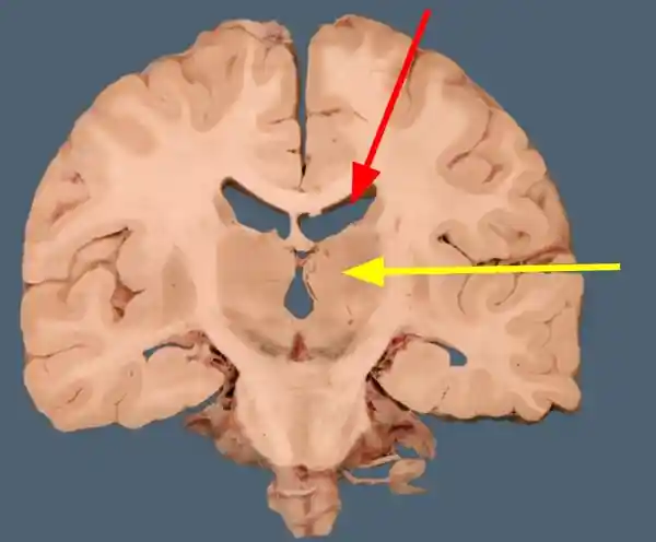
Sample Neuroanatomy Questions:
1. Identify the ventricle at the red arrow 2. Name the nucleus that is immediately lateral to the ventricle at the red arrow 3. Identify the structure at the yellow arrow
Short Sample Patient Diagnosis
A patient comes into your office with recent developments of shakes and tremors. You notice that their hands randomly jut out and they have a lot of twitching in their face. The patient has nothing visibly wrong with them (i.e. no cuts, no infections, etc.) and mention that it all started with slight hand twitches. You get them a genetic test and it comes back with a mutation in the htt gene. Based on this description, what is this patient most likely suffering from?Bipolar Disorder, 7. Huntington's Disease, 8. Insomnia, 9. Multiple Sclerosis, 10. Narcolepsy, 11. Parkinsons Disease, 12. Prosopagnosia, 13. Schizophrenia, 14. Stroke, 15. Tourette's Syndrome
Final Showdown for Top 3 Competitors
Live Q&A
Top 3 Competitors will rotate through each of the 3 rounds individually. Live Q&A will take place in the lecture hall where the emcee will read aloud 10 questions for the competitor to answer orally. The other 2 competitors will be in their own round.
Live Patient Diagnosis
Top 3 Competitors will rotate through each of the 3 rounds individually. Live patient diagnosis will consist of a 3 minute interview with a patient acting out a neurological disorder. You, as the competitor will ask 'yes or no' questions to diagnose the patients condition. and answer 3 follow-up questions.
Neuroanatomy & Neurohistology
Top 3 Competitors will rotate through each of the 3 rounds individually. Neuroanatomy & neurohistology will take place in the lab in a bell ringer style. There will be 5 stations with different parts of the brain labelled along with 3 slides under the microscope to be identified as well as their function. **1 minute will be given per station.